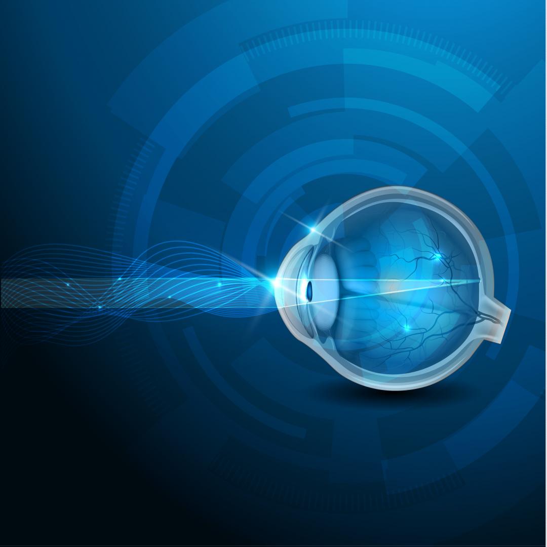
LASER MICROSURGERY

eyerobotica

LASER MICROSURGERY

The Ophthalmological Centre EyeRobotica puts an end in presbyopia and cataract and the problems of daily life they may cause, with the new, state-of-the-art equipment VICTUS Femtosecond Laser Platform of the US company Bausch+Lomb.

It is the most modern invasive surgery for the treatment of presbyopia and cataract, with the ability to correct near, mid-distant or distant vision at the same time with the use of intraocular multifocal lens.

The Laser VICTUS Femtosecond Laser Platform is the only one designed for this type of surgery and ensures:

  • Correction of presbyopia, astigmatism and/or cataract all at once
  • 3D image of the eye
  • Safety during surgery
  • Precision in cutting
  • Less surgical time
  • Less intervening in the eye
  • Faster recovery

The Laser of the Ophthalmological Centre EyeRobotica is fast with the ability to complete the surgery in a few seconds!

This Laser is the cutting edge of Ophthalmology and provides a definite and permanent solution for presbyopia and cataract. It is a bloodless and painless surgery which does not require hospitalization. The patient returns to daily routine right away, without feeling any pain or discomfort.

PHOTOTHERAPEUTIC KERATECTOMY (PTK)

eyerobotica

PHOTOTHERAPEUTIC KERATECTOMY (PTK)

Phototherapeutic Keratectomy is applied as an effective treatment for a variety of superficial corneal disorders. It is an additional use of Excimer Laser, during which removing tissue from the cornea helps the treatment of anterior surface clouding. This clouding is usually a result of an injury, inflammation and/ or of unknown etiology.

The technique demands preoperative research of the patient and assessment of the extent and depth of the clouding.

CORNEAL RINGS INSERTION OF INTRASTROMAL CORNEAL RING SEGMENTS

eyerobotica

CORNEAL RINGS INSERTION OF INTRASTROMAL CORNEAL RING SEGMENTS

CORNEAL RINGS

Corneal rings, also called corneal ring segments, are small plastic devices implanted at the corneal stroma to smooth the shape of the front portion of the eye and to reduce the refractive error, which is due to keratoconus, and other ectatic disorders of the cornea.

Also, they have been designed for the treatment of keratectasia, providing a better result in smoothing the corneal surface.

Technique UV COLLAGEN CROSS-LINKING

eyerobotica

Technique UV COLLAGEN CROSS-LINKING

CXL

In recent years an emerging treatment method has been applied in ophthalmology. Corneal collagen cross-linking is a technique which uses UV-A light and a photosensitizer to strengthen chemical bonds in the cornea.

The goal of the treatment is to halt progressive and irregular changes in corneal shape.

The process starts by removing the epithelium of the cornea, and then instilling drops of riboflavin 0.1% (vitamin B2) for 10 minutes.

This stage of the treatment is to allow riboflavin to diffuse into the cornea.

After adequate riboflavin absorption, the patient is positioned with the UV-A light for 4 more minutes. Total treatment time for each eye is about 15 minutes. In the end, a contact lens is been applied and the patient returns home.

The best candidates for this treatment are patients with diagnosed keratoconus, after a clinical examination and Corneal Topography have been conducted, and also those with other degenerative defects (i.e. peripheral corneal thinning), or patients who are suffering from corneal ectasia after a refractive surgery.

Deterioration and progression of the disorder is perceived by the patient due to the frequent changes of vision glasses or contact lenses. The ophthalmologist detects the condition after conducting a special examination, such as topography and corneal pachymetry.

Not ideal candidates are: people older than 35 with a stable keratoconus, pregnant or breastfeeding women, people with a herpes infection of the eye, or chemical injuries of the cornea or people allergic to riboflavin.

Selecting the appropriate patients and applying the method properly makes the treatment safer and more effective and decreases the possibility of a need for corneal transplant in more than 50%.

PΤK + CXL

Patients with keratoconus who are not able to combine the surgery of stabilizing keratoconus with Laser for correcting another refractive error, can be subjected to Laser to smooth corneal epithelium together with cross- linking.

Laser precedes cross-linking and aims at the improvement of the cone – not full correction-and of aberropia. As for the patient, better vision and stabilization are gained at the same time.

PRK + CXL

Patients with keratoconus in early stages can combine surgery for stabilizing keratoconus with Laser. Laser comes before or after stabilization and aims at partial or full correction of the eye.

As for the patient, better vision and stabilization are gained at the same time. Customizing the patients and even each of the eyes separately is the key to the success of this method.

Technique PRK (PHOTOREFRACTIVE KERATECTOMY)

eyerobotica

Technique PRK (PHOTOREFRACTIVE KERATECTOMY)

The PRK technique is very effective in correcting small degrees of myopia, hyperopia and astigmatism.

In PRK, the surface membrane of the cornea (epithelium) is removed using a special tool, and then Excimer Laser treatment is applied, to modify the cornea curvature and correct the refractive error.

At the end of the surgery, a contact lens is applied to help the epithelium heal within the next three to four days. Full recovery and restoration of vision is slower with this technique compared to LASIK technique, because PRK alters the external surface of the cornea.

PRK is recommended as a method instead of LASIK for patients with thin corneas and for the correction of a rather small refractive error. The removal of the cornea’s epithelial layer is accomplished with a special tool and the help of either the laser or a liquid solution. This is called ‘NO TOUCH’ PRK.

Technique PRK XTRA (PHOTOREFRACTIVE KERATECTOMY EXTRA)

In cases of patients with high degrees of a refractive error and marginal cornea thickness for PRK, we apply the technique PRK XTRA. Through this procedure we can correct ametropia and at the same time stabilize the cornea and eliminate the improbable case of postoperative ectasia.

Thus, after applying the classic technique PRK, and before placing the contact lens, we instill riboflavin drops for 90 seconds and then irradiate the cornea with ultraviolet light for 90 more seconds.

Technique LASIK (LASER IN SITU KERATOMILEUSIS)

eyerobotica

Technique LASIK (LASER IN SITU KERATOMILEUSIS)

LASIK technique is a surgical procedure which is performed in two steps. First, a fine corneal flap is lifted by applying a special laser. Then, another piece of equipment, the Excimer Laser, modifies the corneal curvature in order to correct the refractive error.

The corneal flap is attached back to its original position, without requiring any stitches. This process lasts only a few minutes.

Ophthalmic drops are instilled into the eyes to facilitate the process of treatment.

Visual restoration is significantly fast and there is little to no postoperative discomfort. The replacement of the classic microkeratoms with new Laser microkeratoms led to the use of the international term “Femto LASIK”.


Technique LASIK XTRA (LASER IN SITU KERATOMILEUSIS EXTRA)

In cases of patients with high myopia or astigmatism and marginal cornea thickness to apply the technique LASIK, we use the technique LASIK XTRA. Through this procedure we can correct ametropia and at the same time stabilize the cornea and eliminate the improbable case of postoperative ectasia.

Thus, after applying the classic technique LASIK, and before repositioning the cornea flap, we instill riboflavin drops for 90 seconds, then attach the flap back to its position and irradiate the cornea with ultraviolet light for 90 more seconds.

Θεσσαλονίκη Ανατολικά

Θεσσαλονίκη Δυτικά